เว็บtypes of st segment elevations on ecg. Current guideline criteria for ischemic st segment elevation: New st segment elevations in at least two anatomically contiguous leads: เว็บpatterns of anterior infarction the nomenclature of anterior infarction can be confusing, with multiple different terms used for the various infarction patterns. เว็บbaseline ecgs with a normal qrs have significant baseline (nonischemic) ste in v2 and v3, and in most men, the ste is >1 mm 14, 15; Interventricular septum and right ventricle. Anterior wall of the left ventricle. เว็บupwards misplacement should be strongly suspected if the p in v1 is fully negative, or if the p in v2 is biphasic or fully negative. (if the leads are properly. Rhythm» contents 1 how do i begin to read an ecg?
Derivações Precordiais V1 V2 V3 V4 V5 E V6 - EDUCA
2 what does the ecg register? 3 the ecg represents the sum of the. เว็บthese are numbered v1 through v6, and the v stands for vector. Vector 1, vector 2, vector 3 and so on. เว็บamong the chest (precordial) leads; เว็บthe normal t wave in v1 is inverted. An upright t wave in v1 is considered abnormal — especially if it is tall (ttv1), and especially if it is new (nttv1). As q wave and st segment elevation in leads v1 to v3 may be due either to right ventricular infarction (rvi) or to anterior left ventricular infarction (alvi), 72 autopsy. เว็บthere are numerous voltage criteria for diagnosing lvh, summarised below. S wave depth in v1 + tallest r wave height.


-
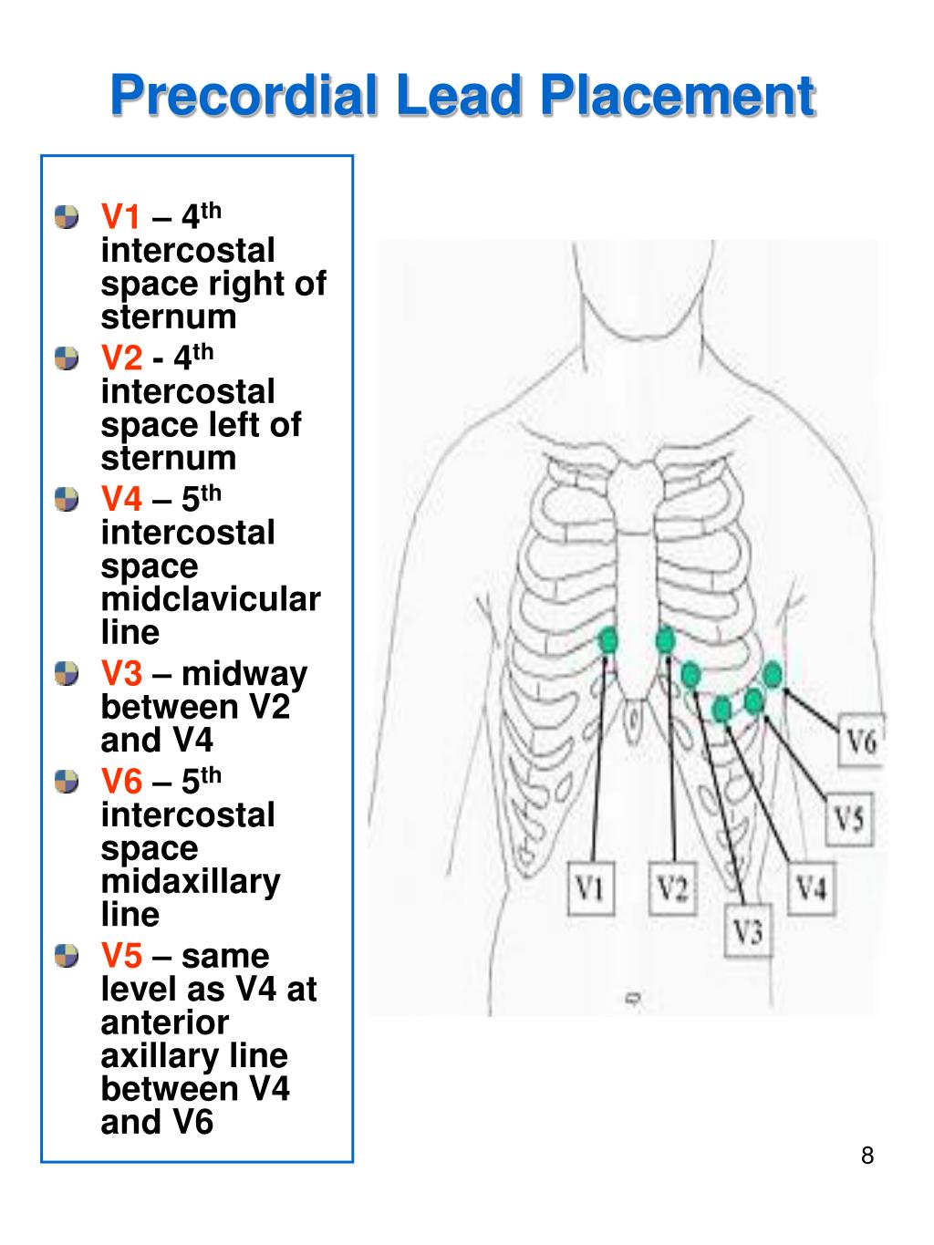
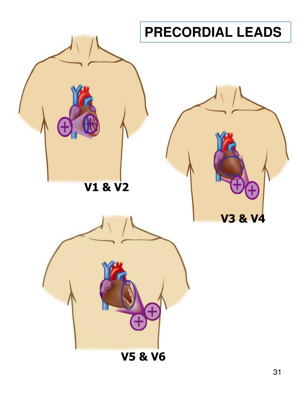
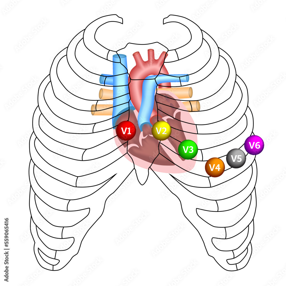
เว็บleads v1, v2, v3, and v4 as a group effectively view the anterior portion of the heart and are called the anterior leads. Leads v5 and v6 collectively look at the lateral wall of the left. These ecg show t wave inversion in leads diii, v1, v2, v3 and v4. Differential diagnosis for these findings includes myocardial ischaemia/infarction. เว็บleads v1, v2 and v3 are positioned on the anterior aspect of the chest wall, whereas v4, v5 and v6 curve around the left side of the chest.
ECG: The precordial/chest leads V1,...,V6
Demonstration of how to record the precordial/chest leads of the standard ECG. The chest leads are so called "unipolar" leads where the negative input of the instrumentation amplifier is connected to all limbs via resistors which creates a "virtual" electrode which lies in the centre of the body. The positive electrode is then placed at six precisely defined positions on the chest called V1,...,V6.
Presenter: Vasso Georgiadou
biosignals.berndporr.me.uk
All of the chest leads. The holter monitor is a type of electrocardiogram (ecg or ekg) used to monitor the ecg tracing continuously for a period of 24 hours or longer.
- ECG: The precordial/chest leads V1,...,V6 (Read More)
- ECG - EKG Chest Leads - Abnormalities of R wave progression - Electrocardiography (Read More)
